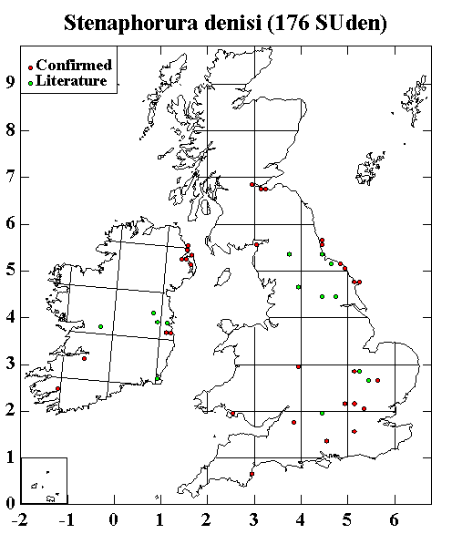
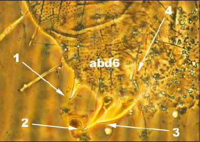
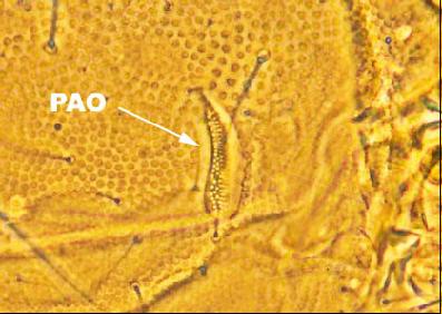
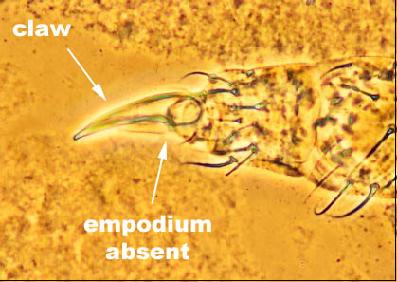
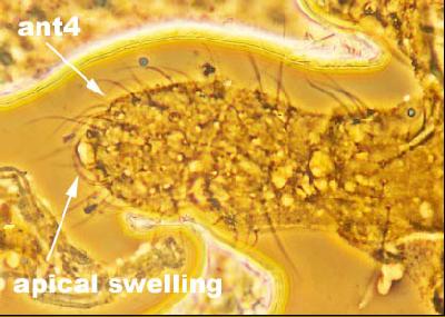
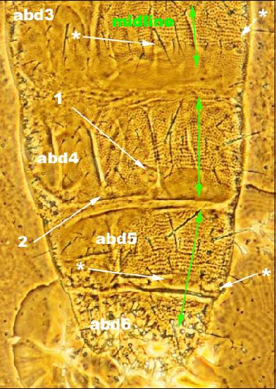
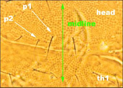
Species of Stenaphorura are white and blind, possess four anal spines (Fig. 1), and have an elongate post-antennal organ (PAO) with 70 or more small vesicles (Fig. 2). The foot does not possess an empodium (Fig. 3). Stenaphorura denisi (up to 1.5 mm in length) was described by Bagnall (1935) and, together with Stenaphorura lubbocki, has been considered by some authors as a junior synonym of Stenaphorura quadrispina. However, following examination of all the Stenaphorura slides in the NHML, I am convinced that all three are distinct species. About 15% of the specimens were incorrectly identified so all earlier records of Stenaphorura cannot be uncritically accepted unless they are supported by specimens. The most easily observed characters that distinguish Stenaphorura denisi are presence of an apical swelling on the tip of the antenna (ant4) (Fig. 4), a pair of bumps on the sixth abdominal segment (abd6) (Fig. 1), and head with setae p1 longer than p2 (Fig. 5). There are also two dorsal pseudocelli on each side of the fourth abdominal segment (Fig. 6). Stenaphorura denisi appears to be widely distributed throughout the UK and Ireland
Fig. 1 (above): Sixth abdominal segment (abd6) of Stenaphorura denisi collected from Culross, Fife, Scotland in May 1937 by Bagnall. There are four anal spines (1-4). There is also a small bump on each side just anterior to anal spines 1 and 4.
Fig. 2 (above): Post-antennal organ (PAO) of a paratype of Stenaphorura denisi collected from Corstophine near Edinburgh in February 1935 by Bagnall.
Fig. 3 (above): Foot of the third leg of the same specimen of Stenaphorura denisi shown in Fig. 1.
Fig. 4 (above): Antenna of the holotype of Stenaphorura denisi collected from Speeton, E. Yorks. in September 1934 by Bagnall. Note the apical swelling on the fourth antennal segment (ant4).
Fig. 5 (above): Posterior margin of the dorsal side of the head of the holotype of Stenaphorura denisi collected from Speeton, E. Yorks. in September 1934 by Bagnall. The p1 setae are longer than the p2 setae. th1, first thoracic segment.
Fig. 6 (above): Dorsal side of the posterior abdomen of Stenaphorura denisi collected from Newmarket, Suffolk in March 1971 by A. Baker. The third (abd3) and fifth (abd5) segments have only one pseudocellus (PSO) on each side (*) but there are two PSO on each side of the fourth abdominal segment (abd4).