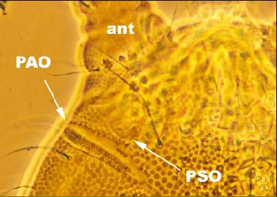
Species of Paratullbergia are relatively large for members of the subfamily Tullbergiinae (up to 1.4 mm in length). They are soil-dwelling Collembola and have particularly prominent anal spines as long as a foot claw (Fig. 1). The pseudocelli do not have the characteristic rosette shape of Mesaphorura species (Figs. 2 and 3). The post-antennal organ is long and thin with about 50 vesicles arranged in two rows (Figs. 4 and 5). There is no empodium on the foot. Two species occur in UK/Eire. Paratullbergia callipygos is by far the most common and is widely distributed. Paratullbergia macdougalli is much less common but its chaetotaxy is distinctive from Paratullbergia callipygos. Well-oriented and cleared adult specimens are easy to identify under phase contrast illumination. On the dorsal side of the second (abd2) and third (abd3) abdominal segments of Paratullbergia callipygos, the setae a2-a1-a1-a2 are in a straight line (Fig. 6) whereas in Paratullbergia macdougalli, setae a1-a1 are distinctly further forwards than the a2 setae. Also in Paratullbergia callipygos, there is a pair of tubercles posterior to the crescent-shaped ridges (Fig. 7); these tubercles are absent from Paratullbergia macdougalli. Many of the literature records of Paratullbergia callipygos are of the junior synonym Paratullbergia carpenteri. I have also examined the holotype of Paratullbergia. concolor described by Womersley (1930) from Epping Forest and it is clearly a junior synonym of Paratullbergia callipygos (features of the original description do not match those of the specimen).
Fig. 1 (above): Composite photograph of Paratullbergia callipygos of 1.2 mm in length collected from Lightwater, Surrey in May 1951 by T. Clay and J.T. Salmon.
Fig. 3 (above): Pseudocellus of the same specimen of Paratullbergia callipygos shown in Fig. 1.
Fig. 2 (above): Dorsal side of the fourth abdominal segment (abd4) of the same specimen of Paratullbergia callipygos shown in Fig. 1. PSO, pseudocellus.
Fig. 5 (above): Post-antennal organ (PAO) of the same specimen of Paratullbergia callipygos shown in Fig. 1.
Fig. 4 (above): Left side of the head of the same specimen of Paratullbergia callipygos shown in Fig. 1. ant, antenna; PAO, post-antennal organ; PSO, pseudocellus.
Fig. 7 (above): Dorsal side of the sixth abdominal segment of the same specimen of Paratullbergia callipygos shown in Fig. 1. cr, crescent-shaped ridges; t, tubercles.
Fig. 6 (above): Dorsal side of the third abdominal segment (abd3) of the same specimen of Paratullbergia callipygos shown in Fig. 1. Setae a2-a1-a1-a2 are in a straight line.
Fig. 8 (above): Female genital plate (fgp) on the ventral side of the fifth abdominal segment (abd5) of the same specimen of Paratullbergia callipygos shown in Fig. 1. Ant, anterior; P, posterior.